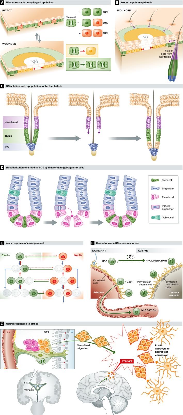
Figure 3. Stem cell responses to stress.
(A) Wound repair in oesophageal epithelium. Following wounding, progenitor cells next to the wound (w) exit the cell cycle and migrate towards the defect (yellow area and inset). Behind this ‘migrating front’, cycling progenitors undergo divisions heavily biased to self-duplication (green area and inset), expanding the progenitor compartment to generate the excess cells required to repair the epithelium. Once the wound has closed, progenitor behaviour switches back to homoeostasis. (B) Wound repair in epidermis. In the epidermis, progenitor cells change their behaviour as in the oesophagus, but wound repair is supported by a flux of cells into the epidermis from hair follicles (purple arrows) and, in tail skin, mobilisation of quiescent reserve cells in the epidermis. Migration explains the appearance of radial clones around a healed wound seen in lineage-tracing experiments. (C) HF ablation and repopulation. Following ablation of SCs in the bulge (green) of telogen HFs, cells in the upper follicle (purple) migrate and proliferate (purple arrows) to reconstitute the bulge SC population, enabling the hair cycle to proceed normally. (D) Reconstitution of intestinal SCs by differentiating progenitor cells. Following ablation of Lgr5-expressing SCs in the crypt base (green), Paneth cell precursors (blue with pink border) dedifferentiate and colonise the crypt base niche with SCs functionally equivalent to the original population. (E) Injury response of male germ SCs. Following treatment with the cytotoxic agent busulphan, the GFRα1+ SC population is depleted (grey outlines) but then reconstituted by surviving Ngn3+ cells reverting into GFRα1+ cells (green arrows) and a decrease in the probability of GFRα1+ cells transferring to the Ngn3+ compartment (pink dotted arrows). (F) Haematopoietic SC stress responses. Dormant SCs are recruited into cycle by treatment after damage to the haematopoietic system from a cytotoxic drug (5FU) or cytokines, such as Gcsf, released after infection. Gcsf also triggers dormant SCs to migrate into the circulation. (G) Neural SC responses to stroke. Following a CNS stroke, which results in cell death due to ischaemia, clusters of neuroblasts (orange) are seen in the injured region. Lineage tracing argues these derive both from mobilisation of neural SCs in the SVC followed by migration to the site of injury (orange with green border) and from differentiated astrocytes within the area of the stroke entering a neurogenic programme (orange with red borders).

