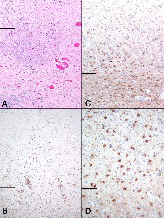
Figure 3. Area of HCS in a PML patient with idiopathic CD4+ lymphocytopenia.
Horizontal lines separate cortex (above) from white matter (below) (A) Myelin stain demonstrates nearly complete demyelination with rare relatively preserved areas (LFB-H&E, 100x). (B) CD68 staining shows perivascular and interstitial macrophages and activated microglia present in white matter as well as deep cortex (CD68, 100x) GFAP staining at low and high magnifications (C and D) highlights prominent astrogliosis in white matter and JCV focal leukocortical encephalitis in deeper cortical layers (C-100x, D-400x).

