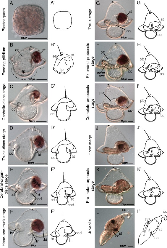
Figure 2.

Development of Micrura alaskensis. Differential interference contrast images. Lateral views except in (A). Apical organ (indicated by asterisk) orientated up, juvenile anterior to the left. (A) Blastosquare stage. Polar view (animal or vegetal). (B) Feeding pilidium stage larva. Esophagus (es), leads to blind stomach (st), which is dark red due to algal food. Ciliated band, indicated with dashed line, spans lobes and lappets (lp). (C) Cephalic-discs-stage larva. One of the paired cephalic discs (cd) labeled. (D) Trunk-discs-stage larva. One of the paired trunk discs (td) labeled. (E) Cerebral-organ-discs-stage larva. One of the paired cerebral-organ discs (cod) labeled. (F) Head-and-trunk-stage larva. Cephalic discs have merged with each other to become the head rudiment (hr). Cerebral-organ discs have merged with the trunk discs to become the trunk rudiment (tr). Dorsal disc (dd) is present. (G) Torus-stage larva. All discs have merged together to form toroid of juvenile tissue around larval stomach. Cerebral organ (co) is labeled. (H) Extended-proboscis-stage larva. Proboscis (pb) has begun to grow over the esophagus towards the stomach. Caudal cirrus (cc) is evident. Membrane housing juvenile (the amnion) is decorated with red-brown pigment spots in a polka-dot pattern. Amniotic pigment spots develop in earlier stages, but are quite clear in this image. (I) Complete-proboscis-stage larva. Proboscis has grown to meet stomach. (J) Hood-stage larva. Dorsal tissues of juvenile overgrow proboscis. (K) Pre-metamorphosis-stage larva. Juvenile is complete inside larva. Metamorphosis (not shown) is rapid and radical; juvenile breaks out of larval body and devours all larval tissues in a minute or less. (L) Juvenile after metamorphosis. Larval structures, including amniotic pigment, are inside juvenile stomach.
