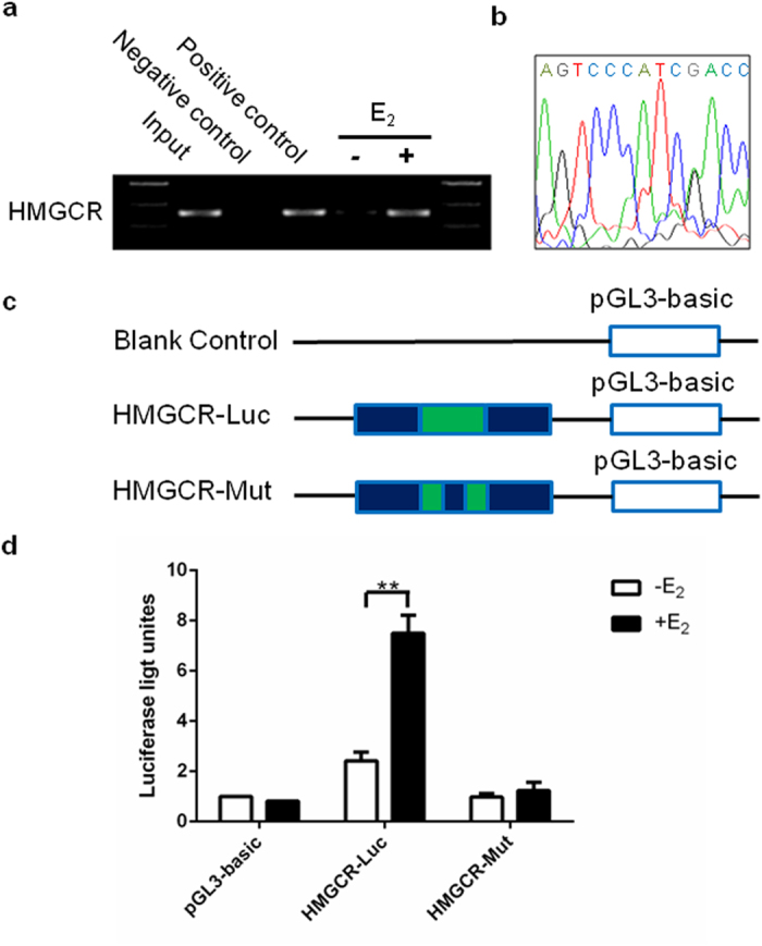
Figure 4. A functional ERE was identified within the HMGCR promoter.

(a) ChIP analysis was performed using anti-ERα or anti-RNA Polymerase II antibody to ascertain the existence of the ERE in the promoter of the HMGCR gene. The PCR results showed that one fragment containing the putative ERE could be precipitated after treatment of HepG2 with E2 (10−7 mol/L) for 24 hours. (b) The pulled down band was excised from the gel and sequenced. (c) Schematic diagram of luciferase reporter constructs. Blank control: pGL3-basic plasmid; HMGCR-Luc: pGL3-basic plasmid with the putative ERE-like sequence insert; HMGCR-Mut: pGL3-basic plasmid with the mutative ERE-like sequence insert. (d) Luciferase activities of three report systems in the presence or absence of E2 (10−7 mol/L) were compared with each other. The experiments were repeated three times and data were presented as Means ± SEM. **P < 0.01 compared with the value in non-E2 treated control group. ERE: estrogen response element; HMGCR: 3-hydroxy-3-methylglutaryl-CoA reductase; ChIP: chromatin immunoprecipitation; E2: estradiol.
