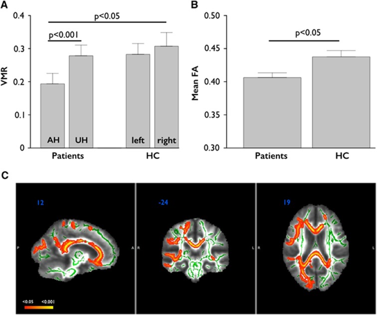
Figure 1.
Differences between groups regarding vasomotor reactivity (VMR) and mean fractional anisotropy (FA). (A) Differences in VMR between hemispheres in patients and healthy controls (HC). (B) Differences in whole-brain mean FA between patients and HC. (C) Differences between groups regarding FA. The side of stenosis/occlusion in patients was set to the right. Patients showed diffuse and significant FA decreases (red-yellow; P<0.05, corrected using threshold-free cluster enhancement (TFCE); white matter skeleton shown in green) when compared with HC, particularly in the frontoparietal regions ipsilateral to the stenosis, including anterior thalamic radiation, forceps minor, forceps major, and corpus callosum. Data in (A and B) are given as mean±s.e.m. AH, affected hemisphere; FA, fractional anisotropy; LH, left hemisphere; RH, right hemisphere; UH, unaffected hemisphere.

