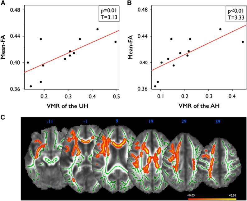
Figure 3.
Correlations between vasomotor reactivity (VMR ) and fractional anisotropy (FA) in patients. (A) Correlations between mean FA of the unaffected hemisphere (UH) and VMR of the UH in patients, and (B) correlations between mean FA of the affected hemisphere (AH) and VMR of the AH in patients. (C) Clusters of FA that are significantly correlated with VMR of the AH in patients (red-yellow; white matter skeleton shown in green). Colors indicate significant voxels (P<0.05, corrected using TFCE), superimposed on a study-specific FA template.

