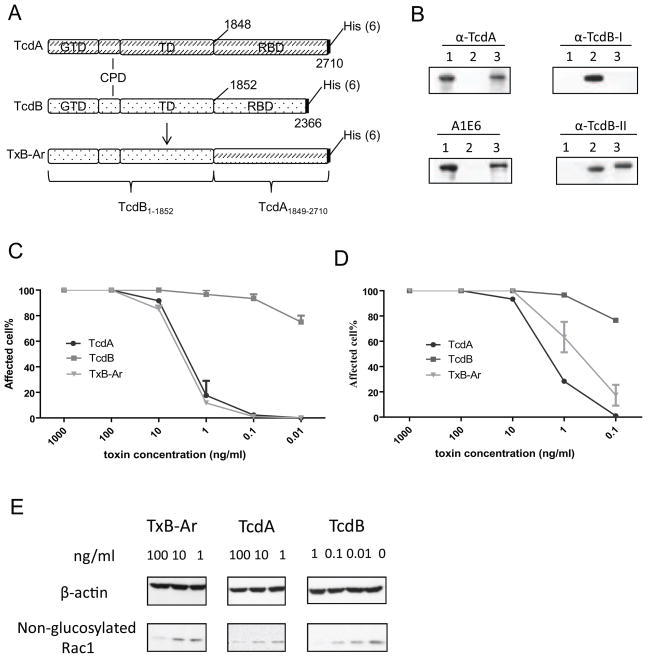
Fig. 1.
Structure and activities of the chimera TxB-Ar. (A) Domain structure of TxB-Ar. TxB-Ar is TcdB with its the C-terminus CROPs replaced by the full-length CROPs from TcdA. (B) Western blot analysis of TxB-Ar. Western blot was performed to detect the domains of TcdA, TcdB, and TxB-Ar with various antibodies. Lane 1: TcdA, Lane 2: TcdB, and Lane 3: TxB-Ar. (C, D): Cytopathic effects of the chimera TxB-Ar, TcdA, or TcdB to Vero cells (C) or CT26 cells (D). The serially diluted toxins were applied to the sub-confluent cells for 16 hours. The percentage of rounding cells was examined under a phase contrast microscope. The experiments were performed three times and error bars indicate the standard error of mean (SEM). (E) Vero cells were incubated with the indicated doses of the toxins for 4 hours. Cells were collected and lysed by SDS sampling buffer for western blot to detect Rac1 glucosylation.

