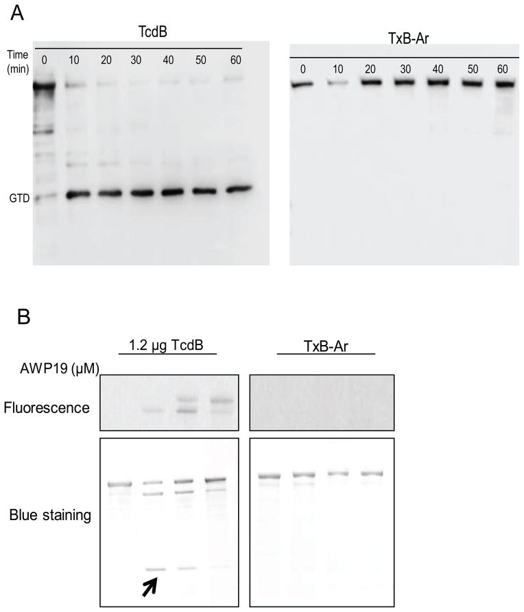
Fig. 2.
Autoprocessing of chimera TxB-Ar. (A) InsP6 induced autoprocessing of TcdB and TxB-Ar. 10 ng/μL of TcdB or TxB-Ar was incubated in the reaction buffer containing 25 μM InsP6 at 37 °C for the indicated time. The reaction was terminated by SDS-sampling buffer and heating at 95 °C for 5 min. A VHH antibody (E3) against the GTD of TcdB was used for western blot analysis. (B) CPD conformational change probed by fluorescent AWP19. TcdB (0.1 μg/μL) was incubated with the indicated doses of AWP19 in 20 μM pH8.0 Tris buffer containing 25 μM InsP6 at 37 °C for 1 hour. The reactions were stopped by SDS sampling buffer and the samples were loaded on a SDS-PAGE. Fluorescence was measured using G-Box Chemi system and the total proteins were visualized on the gel after coomassie blue staining. The arrow indicates the cleaved GTD fragment.

