
Fig. 2.
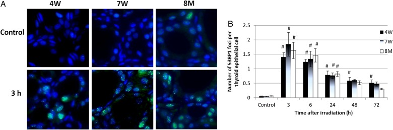
Immunofluorescent staining of p53 binding protein 1 (53BP1) in non-irradiated (control) and irradiated 4W, 7W and 8M thyroid follicular epithelial cells ( × 1000 magnification) (A), and the average number of 53BP1 foci per thyroid epithelial cell after irradiation (B). Data are presented as the mean ± standard error of the mean of the results for three to seven rats per datapoint. # indicates P < 0.05 compared with cells of non-irradiated rats.