Fig. 2.
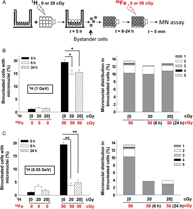
Proton-irradiation propagates protective bystander effects. (A) Experiment schematic: Proton-irradiated cells are plated on the top side of the porous membrane of a Transwell® insert, with bystander cells growing on its underside. Following ∼5 h of co-culture, the bystander cells were harvested and seeded in tissue culture flasks. The bystander cells were exposed to iron ions at different times after seeding. (B) Micronucleus formation (left panel) and micronuclei distribution (right panel) in bystander AG1522 cells that had been in co-culture with cells irradiated with 0 or 20 cGy (shown in brackets) from 1-GeV protons (1H). Bystander cells were exposed 0, 6 or 24 h later to 0 or 50 cGy from 1-GeV/u iron ions (56Fe). (C) Micronucleus formation (left panel) and micronuclei distribution (right panel) in bystander AG1522 cells that had been in co-culture with cells irradiated with 0 or 20 cGy (shown in brackets) from 0.05-GeV protons (1H). Bystander cells were exposed 0, 6 or 24 h later to 0 or 50 cGy from 1-GeV/u iron ions (56Fe). The data show that bystander cells that were co-cultured with low-dose proton-irradiated cells are protected from DNA damage induced by a subsequent challenge of energetic iron ions. *P < 0.05, **P < 0.0001.

