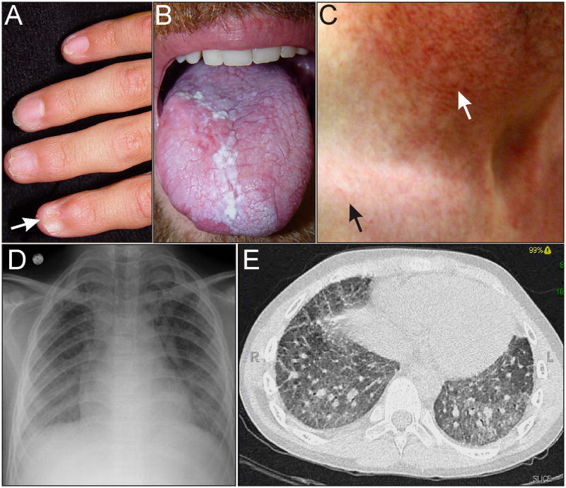
Figure 2.
Phenotypic features of DC. (A) Nail dystrophy (arrow) in an adolescent. (B) Oral leukoplakia in an adult. (C) Skin changes in an adult. Hypopigmented lesions and the typical reticular rash of DC are evident on the upper chest (black arrow). Hypopigmentation is more pronounced in the sun-damaged skin of the neck (white arrow). (D,E) Pulmonary fibrosis on chest radiograph and CT scan of an adult. Photographs courtesy of Susan J. Bayliss, M.D. and William H. McAlister, M.D.

