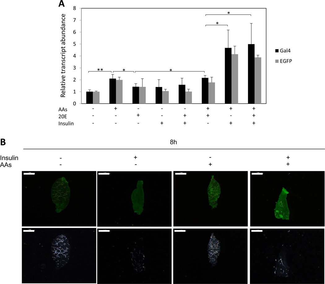
Figure 5.
Effects of AAs, insulin and 20E on expression of the CP-Gal4>UAS-EGFP transgene. (A) Midguts were isolated from non-blood fed CP-Gal4>UAS-EGFP female mosquitoes and incubated under different indicated conditions in vitro for 8 h. Levels of the CP-Gal4>UAS-EGFP transgene transcript were monitored using Gal4 and EGFP primers (Table S3) by means of qRT-PCR. Control transcript level was set at 1, while transcript levels for other treatments were expressed relative to the control. Values are the means of three replicates (± SEM). The experiment was repeated three times. * Indicates statistical significance <0.05. ** Indicates statistical significance <0.01. (B) Detection of EGFP protein in midguts isolated from non-blood fed CP-Gal4>UAS-EGFP female mosquitoes by means of fluorescent immunocytochemistry. The midguts were cultured for 8h under indicated conditions. Immunocytochemistry was performed using the anti- EGFP antibody, followed by incubation with an anti-rabbit fluorescent secondary antibody. Images were obtained using Leica M165FC fluorescent stereomicroscope using GFP-B filter or transparent filter (White light) and LAS V4.0 software. Scale bar: 1mm.

