Fig. 2.

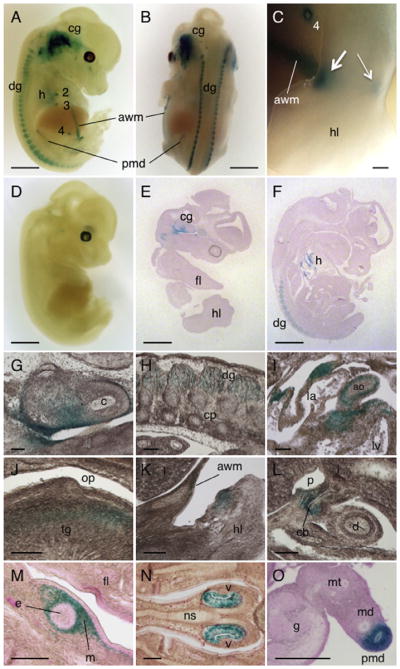
Expression of Dact1-LacZ in embryos. (A–D) E12.5 embryos stained for LacZ activity and photographed as whole mount preparations. (A and B) Dact1LacZ Tg mouse embryos showing strong LacZ signals in the cranial ganglion, dorsal root ganglion, heart, nipples, paramesonephric duct, and abdominal wall mesenchyme. (C) An enlarged view of the hindlimb showing LacZ signals in the ankle with a stronger signal in anterior (thick arrow) than posterior region (thin arrow). (D) WT embryo serving as a negative control. (E and F) Sagittal sections of X-gal-stained E12.5 Dact1LacZ Tg mouse embryos showing strong LacZ signals in the cranial ganglion, heart and dorsal root ganglion. (G–O) Enlarged views of embryonic tissues prepared from E12.5 (G–L) and E14.5 (M–O) Dact1LacZ Tg mouse embryos stained for LacZ activity using X-gal, followed by sectioning. Dact1-LacZ expression was detected in the inner ear (G) and the dorsal root ganglion (H). In the heart, Dact1-LacZ was expressed in the vessel wall and myocardium in the atrioventricular junction (I). Dact1-LacZ was also detected in the intrinsic muscle of the tongue (J) and mesenchyme surrounding some developing structures such as the limb (K), common bile duct (L) and nipple (M). The vomeronasal organ (N) and paramesonephric duct (O) also expressed Dact1-LacZ. cg, cranial ganglion; dg, dorsal root ganglion; h, heart; 2–4, mammary placodes 2–4; awm, abdominal wall mesenchyme; pmd, paramesonephric duct; hl, hindlimb; fl, forelimb; c, cochlea; cp, cartilage primordium; ao, aorta; la, left atrium; lv, left ventricle; op, oropharynx; tg, tongue; l, liver; cb, common bile duct; p, portal vein; d, duodenum; e, epithelial component of mammary placode; m, mesenchymal component of mammary placode; v, vomeronasal organ; ns, nasal septum; md, mesonephric duct; mt, mesonephric tubule; g, gonad (ovary). Scale bars = 1 mm (A, B and D–F), 0.1 mm (C) and 100 μm (G–O).
