Fig. 3.
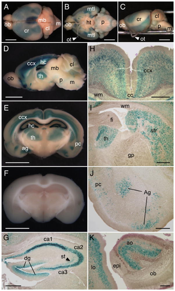
Expression of Dact1-LacZ in adult brain. (A–C) The brains from 8 week-old Dact1LacZ Tg mice were stained for LacZ activity using X-gal and photographed as whole mount preparations. Shown are dorsal (A), ventral (B) and lateral (C) views of the brain. (D–F) The brains from Dact1LacZ Tg mice (D and E) and WT littermates (F) were sliced sagittally along the midline (D) or coronally in the central region of the cerebrum (E and F) and stained for LacZ activity using X-gal. The strongest LacZ signals were detected in the hippocampus, followed by cerebral cortex, ventral forebrain, thalamus and the medial temporal lobes. Weak signals were also noted in the cerebellum, pons and medulla. (G) Within the hippocampus, Dact1-LacZ was detected in the pyramidal neurons in the cornu ammonis regions, granular cells in the dentate gyrus and some interneurons in the stratum radiatum. (H–J) Expression of Dact1-LacZ in other regions of the cerebrum including the cerebral cortex (H), thalamus and striatum (I), amygdaloid nucleus and piriform cortex (J). (K) Expression of Dact1-LacZ in anterior olfactory nucleus and lateral orbital cortex in the ventral forebrain. We analyzed the brains from postnatal day 7 (P7) to 36-week old mice and found similar results. ob, olfactory bulb; cr, cerebrum; mb, midbrain; cl, cerebellum; m, medulla; ht, hypothalamus; p, pons; mtl, medial temporal lobe; ot, optic tract; ccx, cerebral cortex; hc, hippocampus; th, thalamus; ag, amygdaloid nucleus; pc, piriform cortex; ca1–3, cornu ammonis areas 1–3; dg, dentate gyrus; st, stratum radiatum; cc, central canal; wm, white matter; fi, fimbria of hippocampus; gp, external globus pallidus; str, striatum; ao, anterior olfactory nucleus; lo, lateral orbital cortex; epi, accessory olfactory bulb. Scale bars = 3 mm (A–F) and 500 μm (G–K).

