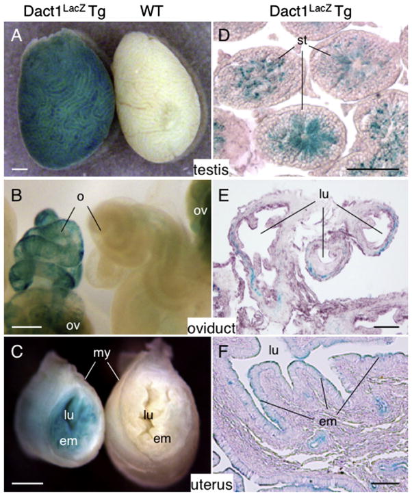
Fig. 7.
Expression of Dact1-LacZ in adult reproductive organs. (A–F) The testis, oviduct and uterus from 8 week-old Dact1LacZ Tg mice and WT littermates were stained for LacZ activity and photographed as whole mount preparations (A–C) or cut into sections, followed by counterstaining with eosin (D–F). Dact1-LacZ was expressed in the late spermatids (D), the connective tissue in the outer wall of the oviduct (E) and the uterine endometrium (F). Note that the ovary shows non-specific LacZ signals (B). We analyzed the tissues from P1 to 35-week old mice and found similar results except for the testes that were negative for LacZ activity before 3 weeks of age. st, seminiferous tubule; o, oviduct; ov, ovary; lu, lumen; em, endometrium; my, myometrial layer. Scale bars = 0.5 mm (A–C), 50 μm (D) and 100 μm (E and F).

