Figure 5.
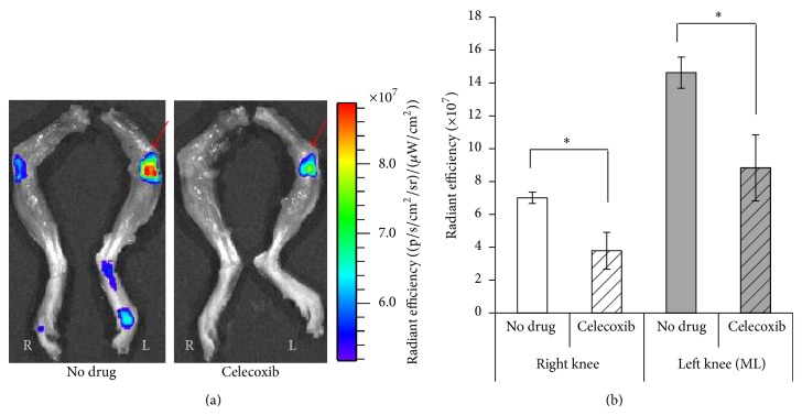
Optical imaging of fluorescently labelled MabCII antibody in PTOA model mice with and without treatment with celecoxib. IVIS scanning shows fluorescent CII targeted antibody binding to the damaged cartilage surface. (a) shows the antibody binding in greater quantity to the loaded left knee in both the drug treated and nondrug treated cases. Antibody binding to the loaded knee of the celecoxib treated group is lower than the loaded knee of the nondrug treated group. The red arrow indicates a mechanically loaded knee. (b) quantifies this binding by measuring fluoresce intensity and calculating radiant efficiency. Results are parallel (a) (n = 6 for each treatment group).

