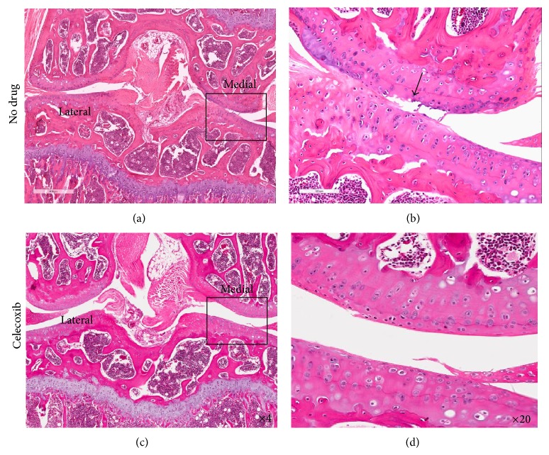
Figure 6.
Histopathology of mechanically loaded knee joints with and without treatment with celecoxib. This figure shows H&E stained coronal sections of loaded mouse knees. Twenty histological sections, each taken 200 μm apart, were analyzed for arthritic joint damage across the entire joint. Sections at the same depth relative to the patella were compared. (a) and (b) are from the mechanically loaded knees of mice receiving no drug treatment (only saline). (c) and (d) are from the mechanically loaded knees of mice treated with celecoxib. In both cases, the lateral tibial and femoral plateaus have minimal damage. The medial plateaus sustained more damage, and thus higher magnification ((b) and (d)) compares treated (d) and untreated medial plateaus (b).

