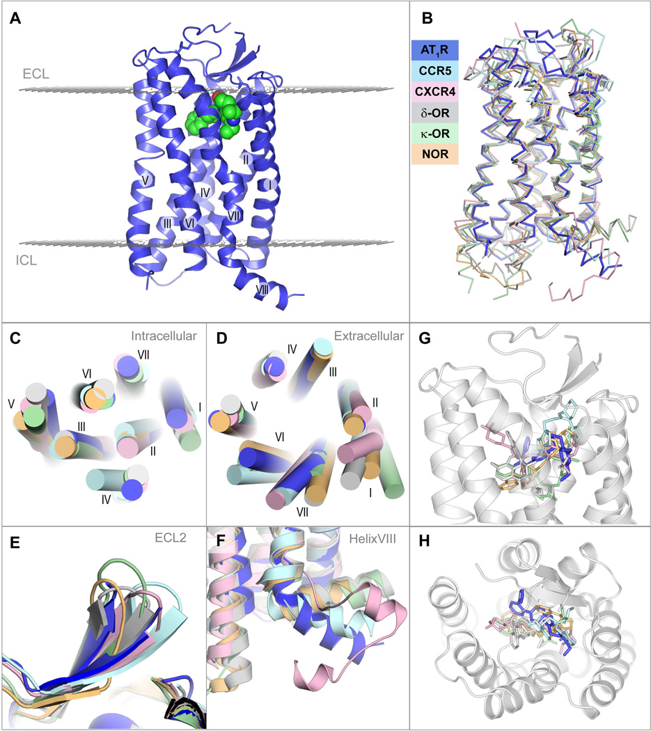
Figure 2. Overview of AT1R-ZD7155 architecture and structural comparison with other peptide GPCRs.
(A) Overall AT1R structure is shown as blue cartoon. ZD7155 is shown as spheres with carbon atoms colored green. Membrane boundaries, as defined by the PPM web server (Lomize et al, 2012), are shown as planes made of gray spheres.
(B) – (G) superposition of AT1R with chemokine and opioid receptors, chemokine CCR5 receptor – light cyan (PDB ID 4MBS), chemokine CXCR4 receptor – light pink (PDB ID 3ODU), δ-opioid receptor – gray (PDB ID 4N6H), κ-opioid receptor – light green (PDB ID 4DJH), NOP receptor – light orange (PDB ID 4EA3), comparing the whole structure (B), intracellular view (C), extracellular view (D), ECL2 (E), helix VIII (F), and the ligand binding pocket side (G) and top (H) views.
See also Figures S1–S2 and Table S1.

