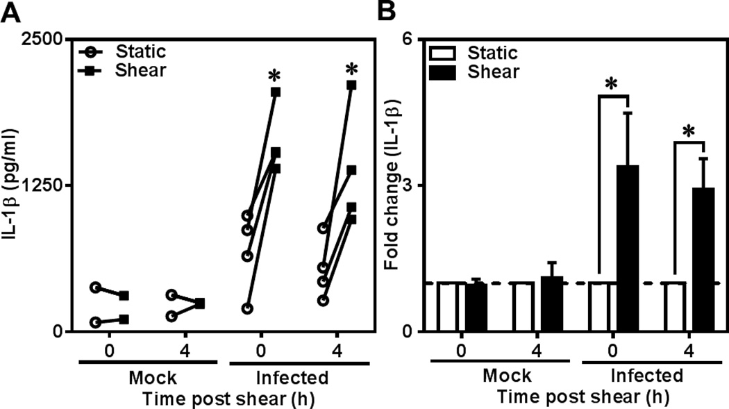
Figure 2.
Effect of shear stress on IL-1β secretion from C. pneumoniae-infected monocytes. Human monocytes were infected with mock (media only) or C. pneumoniae EB (MOI 1) for 2.5 h. 48 h post infection, uninfected and infected cells at a concentration of 6×10⁶ cells/ml were sheared for 1 hour at 0 (static) and 5 dyn/cm2 (shear) using a cone-and-plate viscometer. After shear exposure, the cells were incubated for further 0 or 4 h, and supernatants and cell lysates were analyzed for IL-1β by ELISA. The results are expressed as actual levels (A) and fold increase (B) from one representative experiment performed for four donors and statistical difference was evaluated using two-way ANOVA (*p<0.05).

