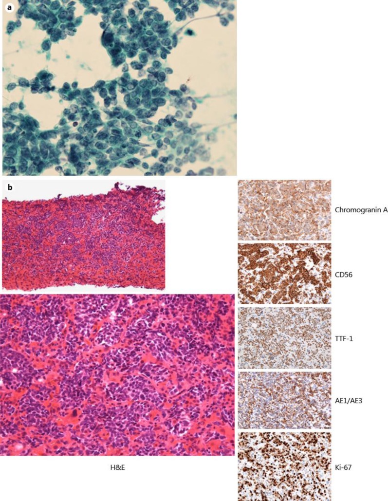
Fig. 3.
a Cytology from a mediastinal lymph node by transbronchial needle aspiration revealed SCLC (×400). b Histological examination of the liver revealed diffuse liver metastases of SCLC, all of which were positive for chromogranin A, synaptophysin, CD56, TTF-1 and AE1/AE3 and with a Ki-67 index of 80% (hematoxylin and eosin: ×40, ×400; chromogranin A, CD56, TTF-1, AE1/AE3, Ki-67: ×400).

