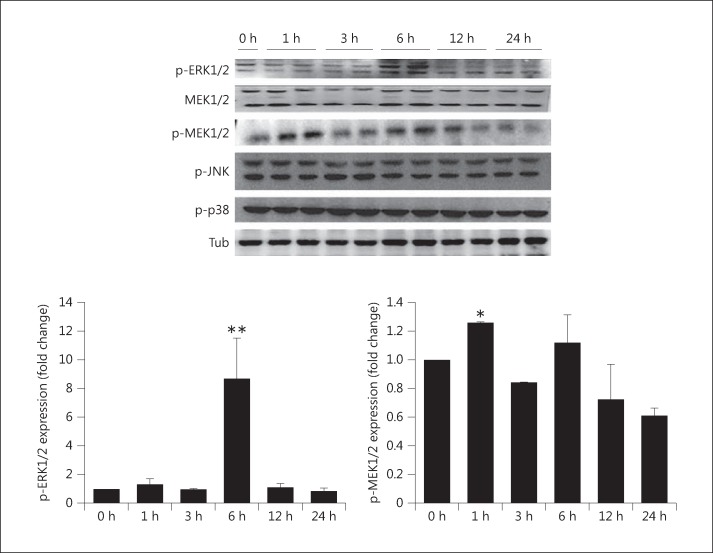
Fig. 4.
Expression of MAPK markers in elevated phosphate-induced myocardial cells with 1.4 mM NaH2PO4. The protein levels of MEK1/2, phosphorylated (p-)ERK1/2, p-MEK1/2, p-JNK, and p-p38 were determined by Western blot analysis. The results are expressed as the mean ± SE values of 3 experiments. * p < 0.05, ** p < 0.01 in comparison to the control. Tub = α-Tubulin.

