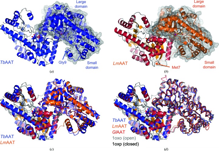
Figure 1.
Open and closed conformations. (a) and (b) show dimers of TbAAT and LmAAT, respectively, in the same orientation. The surface rendition highlights the location of one of the protomers. The locations of the N-termini and the large and the small domains are indicated. The superposition of TbAAT and LmAAT (c) with chain A on the left as a reference highlights the open conformation of TbAAT and the closed conformation of LmAAT. (d) shows an extended superposition of TbAAT, LmAAT and GlAAT, along with open-conformation and closed-conformation chicken AAT (PDB entries 1oxo and 1oxp, respectively). The worm representation highlights the differences for chain B on the right. For simplicity, for chain A on the left only TbAAT-native and LmAAT are shown. This figure was created with CCP4mg (McNicholas et al., 2011 ▶).

