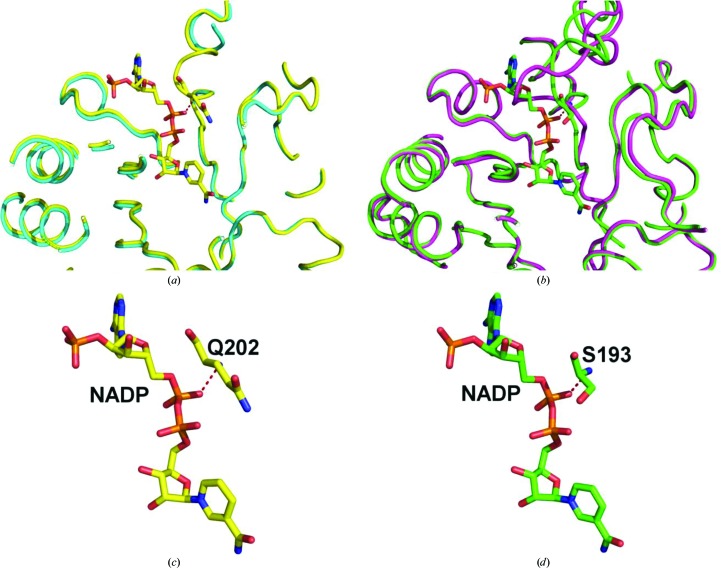
Figure 3.
(a) Overlay of PDB entry 4f40 (blue) and PDB entry 4g5d (yellow) from L. major showing the NADP binding-site area. (b) Overlay of PDB entry 4fzi (purple) and PDB entry 4gie (green) from T. cruzi showing the NADP binding-site area. (c) NADP from PDB entry 4g5d and its interaction with Gln202. (d) NADP from PDB entry 4gie and its interaction with Ser193.

