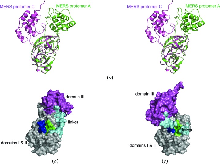
Figure 6.
(a) Stereoview of the superimposed structures of MERS-CoV 3CLpro crystal form III protomer A (green ribbons) and protomer C (magenta ribbons). (b, c) Surface representations of protomer A (b) and protomer C (c) with domains I and II colored gray, the linker loop (residues 188–204) cyan, domain III magenta, the oxyanion loop (residues 143–148) blue and the S1 binding pocket green.

