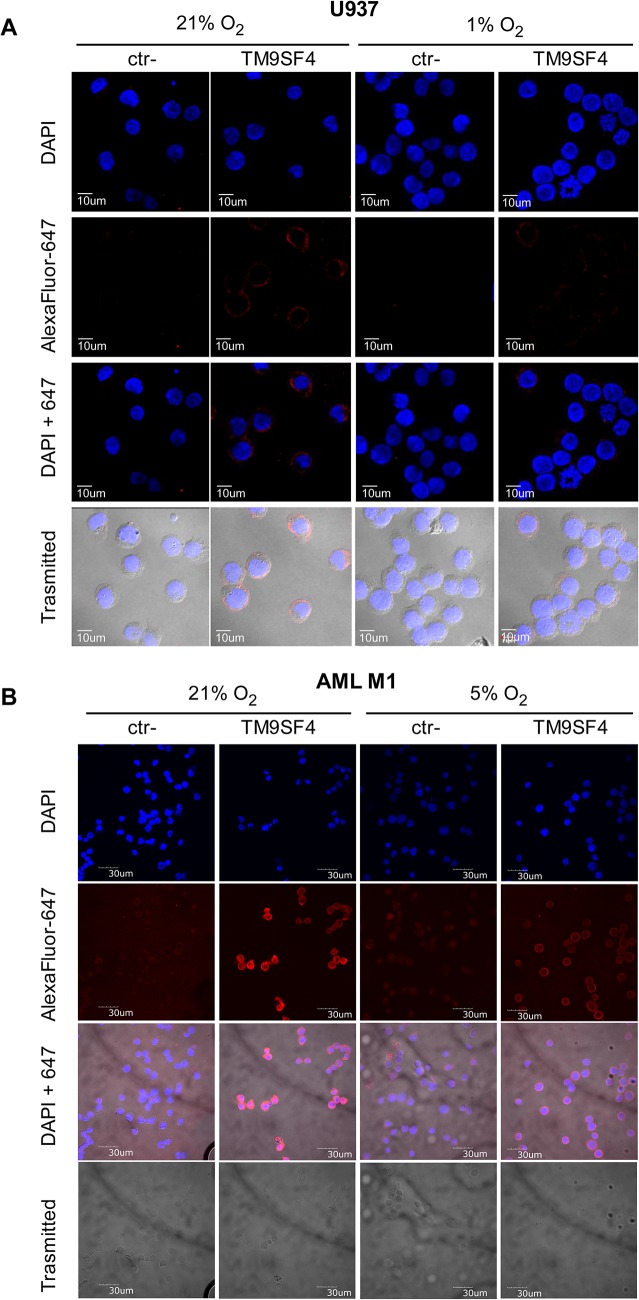
Fig 4. Hypoxia downmodulates TM9SF4 expression in leukemic cells.
(A, B) The decrease of TM9SF4 protein expression in U937 cells (A) and in primary leukemic cells from AML M1 (B) cultured under hypoxic (1% and 5% O2, respectively) compared to normoxic (21% O2) conditions is shown by immunofluorescence analysis of TM9SF4. ctr- is for no primary antibody negative control. DAPI shows nuclear staining; AlexaFluor-647 shows TM9SF4 protein staining; DAPI+647 is the merge of AlexaFluor-647 and DAPI; Trasmitted shows the phase-contrast microscopy fields; Scale bars indicated are 10μm (A) and 30μm (B).

