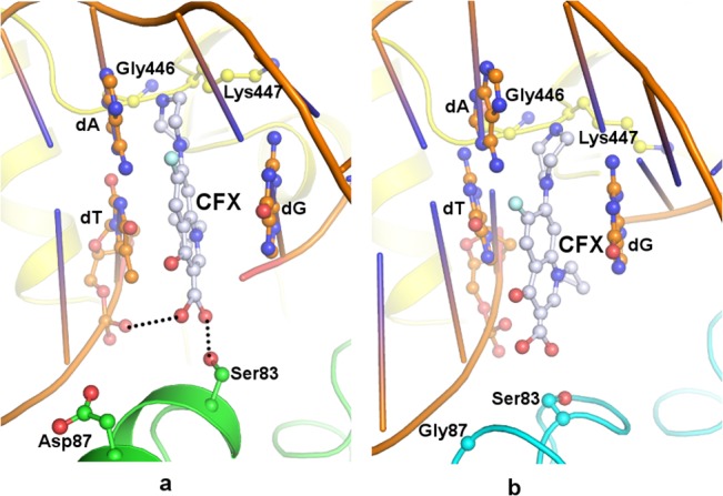
Fig 6. stDNA-Gyrase—ciprofloxacin.

Docked position of ciprofloxacin (ball and stick, grey) in the QBP of stDNA-Gyrase (cartoon) where DNA is drawn in orange, GyrB in yellow (a) wild type GyrA in green and (b) Asp87Gly GyrA mutant in cyan. Ser83 and Asp87/Gly87 are represented in ball and stick in respective colour. Hydrogen bonds are indicated as black dotted lines.
