
Fig 4. 4-1BB-mediated CD25 induction on CD8+ T cells in the presence of PI3K, ERK, or AKT inhibitor.
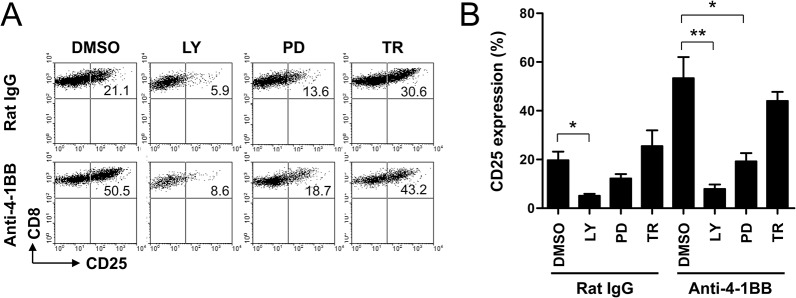
(A-B) Anti-CD3-activated CD8+ T cells for 16 h were pre-incubated with 20 μM LY294002 (LY; PI3K inhibitor), 30 μM PD98059 (PD; ERK inhibitor), or 2 μM Triciribine (TR; AKT inhibitor) for 1 h and then treated with anti-rat IgG or anti-4-1BB mAb for another 24 h. The CD8+ T cells were stained with anti-CD8 and anti-CD25 mAb, and subsequently analyzed by FACSCalibur (BD Bioscience). Live CD8+ T cells were gated and plotted CD8 vs. CD25 (A). Percentages of CD25+ CD8+ T cells were calculated and represented as mean ± SD (n = 5; *, p< 0.05; **, p< 0.01) (B).