
Fig 5. IL-2Rα induction on CD8+ T cells by TNFRSF members.
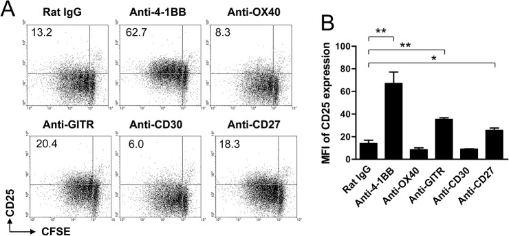
(A-B) CFSE-labeled CD8+ T cells were activated with 0.1 μg/ml of anti-CD3 mAb and simultaneously treated with 5 μg/ml of anti-rat IgG, anti-4-1BB, anti-OX40, anti-GITR, anti-CD30, or anti-CD27 mAb for 3 days. The cells were stained with anti-CD8 and anti-CD25 mAb. CD8-gated cells were plotted as CFSE vs. CD25 (A) and MFIs of CD25 expression were calculated using BD CellQuest software (BD Bioscience) (B). Data are representative of at least three independent experiments. Result in B is mean ± SD (n = 3; *, p< 0.05; **, p< 0.01).