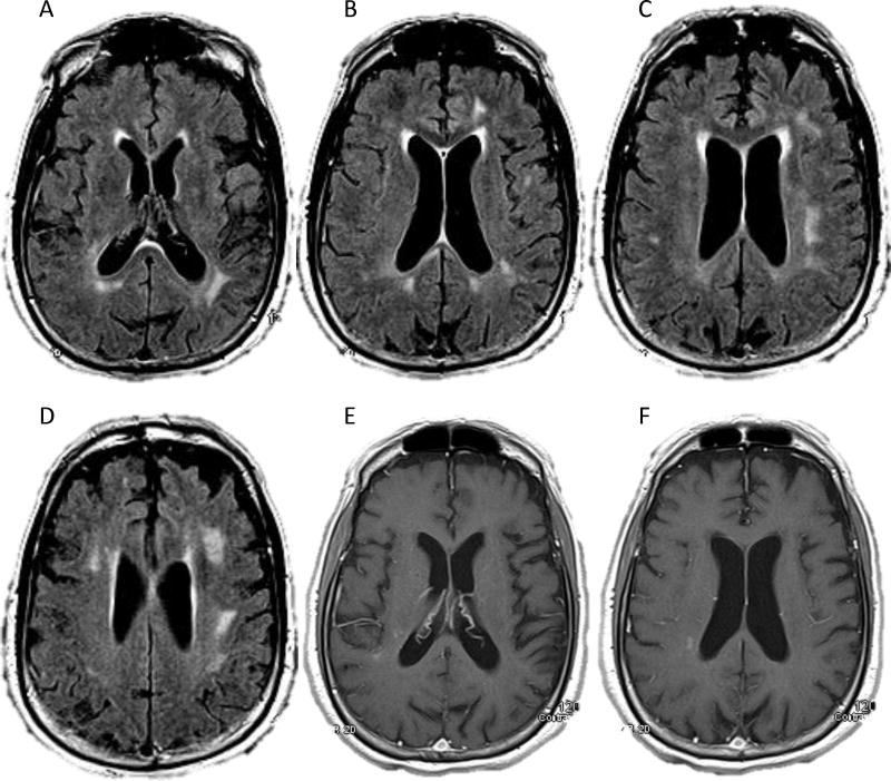
FIGURE 1.
Our evaluation for primary central nervous system lymphoma in a 74-year-old right-handed man with 6 months of progressive personality change, confusion, and impaired gait. On a magnetic resonance imaging scan of his brain, axial FLAIR (Panels A–D) and T1 post-contrast (Panels E–F) sequences showed subcortical and deep periventricular white matter hyperintensities and subtle right periatrial enhancement. Cerebrospinal fluid analysis showed lymphocytic pleocytosis (7 white blood cells/cu mm, 93% lymphocytes), elevated protein (119 mg/dL), normal glucose (52 mg/dL), and negative cytology. Empiric steroids did not improve the patient’s symptoms; however, cerebrospinal fluid analysis while the patient was taking steroids showed resolution of the pleocytosis, persistently elevated protein (96 mg/dL), normal glucose, negative cytology, negative flow cytometry and gene rearrangement from limited specimens, and elevated beta-2 microglobulin (4.6 mg/L). We concluded that the patient had clinically probable lymphomatosis cerebri.

