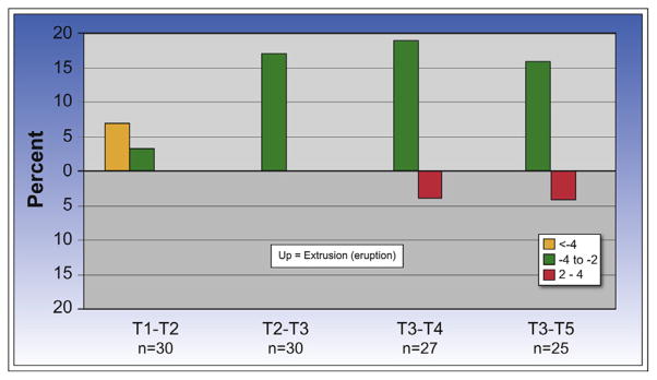
Fig 3.

Percent with change in the mandibular first molar distance from the mandibular plane. During splint therapy (T1–T2), 2 patients had greater than 4 mm of eruption of the mandibular first molars (7%), and 1 (3%) had 2 to 4 mm of eruption. During postintrusion orthodontics (T2–T3), 5 (17%) had 2 to 4 mm of eruption. During the first posttreatment year (T3–T4), 5 (19%) had an eruption of 2 to 4 mm, but 1 patient had an eruption of 2 to 4 mm. During the second posttreatment year, 4 (16%) had 2 to 4 mm of eruption, and 1 had 2 to 4 mm of intrusion.
