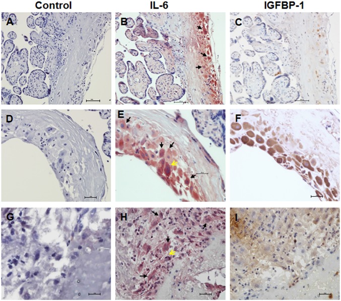
Fig 1. Expression of IL-6 in decidual cells in human placenta.
Paraffin embedded sections of human term placenta (A-F) and frozen sections of idiopathic preterm placenta (G-I) with some attached fetal membrane were analyzed for IL-6 or IGFBP-1 expression by immunohistochemistry. Decidual cells were identified by morphological analysis or IGFBP-1 staining. (A) Term placenta/fetal membrane, control immunostaining. (B) Term placenta/fetal membrane, IL-6 expression is shown in red staining, decidual cells (arrows). (C) Term placenta/fetal membrane, IGFBP-1 expression is shown in brown staining. A-C: adjacent slides of same placenta/fetal membrane. (D) Term placenta/fetal membrane, control immunostaining. (E) Term placenta/fetal membrane, IL-6 expression is shown in red staining, decidual cells (arrows), trophoblast cells (yellow arrows). (F) Term placenta/fetal membrane, IGFBP-1 expression is shown in brown staining. D-F are adjacent slides of another term placenta/fetal membrane at higher magnification. (G) Preterm placenta/fetal membrane, control immunostaining. (H) Preterm placenta/fetal membrane, IL-6 expression is shown in red staining, decidual cells (arrows), trophoblast cells (yellow arrow). (I) Preterm placenta/fetal membrane, IGFBP-1 expression is shown in brown staining. G-I are adjacent slides of idiopathic spontaneous preterm placenta/fetal membrane. At least 3 different placentas were used in each group. Shown in the figure are representative tissue slides. Original magnification: A-C, 20X; D-I, 40X.

