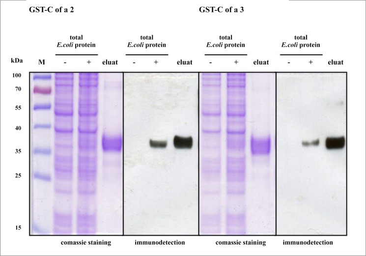
Fig 4. Purification and immunodetection of rCof a 2 and rCof a 3.
Purification of E. coli-expressed rCof a 2 and rCof a 3 under native conditions. Proteins recovered by GST affinity cromatography were separated by SDS-PAGE (eluat). Total E. coli protein fractions before and after IPTG supplement are indicated by − and +; M: molecular wight marker. The first gel stained with coomassie brilliant blue, the second for immunodetection with anti His-Ab. Both gels ran together at same conditions.

