
Figure 6.
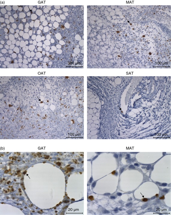
Detection of Neospora caninum in the adipose tissue of infected interleukin-12 (IL-12)/IL-23 p40−/− mice. (a) Representative images showing parasitic forms in the gonadal adipose tissue (GAT), mesenteric adipose tissue (MAT), omental adipose tissue (OAT) and subcutaneous adipose tissue (SAT) of p40−/− C57BL/6 mice 7 days after intraperitoneal administration of 5 × 10⁵ N. caninum tachyzoites, detected by immunohistochemistry. Thin sections of the indicated organs/tissues were specific stained (brown coloration, indicated by arrows) with a polyclonal serum goat anti-N. caninum and counterstained with haematoxylin. This is one representative result of two independent experiments with three mice per experiment. Bar = 100 µm (b) Higher magnification of GAT and MAT showing parasitic forms closely associated with adipocytes (arrow). Bar = 20 µm.