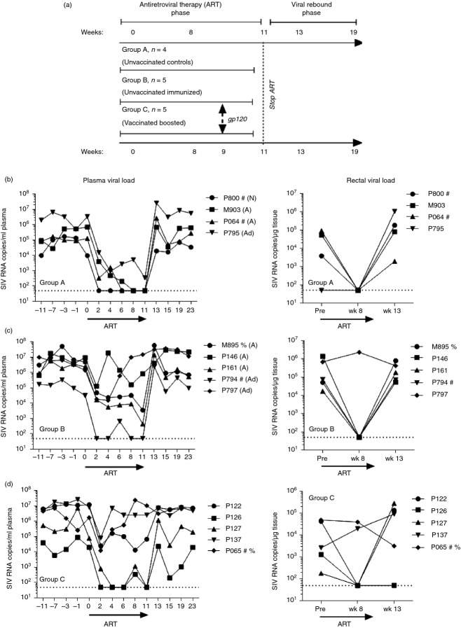
Figure 1.
Study design and plasma and tissue viral loads for individual animals throughout the study. (a) Schematic design of the study. (b–d) Plasma and rectal tissue viral loads were obtained at the indicated time-points by nucleic acid sequence-based amplification (NASBA). Dotted lines indicate the lower limit of viraemia detection (50 copies) by NASBA. Viral loads in Group A [antiretroviral therapy (ART) only controls] were previously reported.26 #, Mamu A*01 positive; %, Mamu B*17 positive; N, naive; A, received alum only; Ad, received empty Ad vector and MPL-SE adjuvant only.

