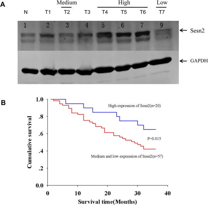
Fig 5. Low levels of Sesn2 expression associate with poor survival in Chinese lung cancer patients.
(A) Tissue homogenates prepared from resected lung cancer tissues were subjected to immuno-blotting probed with the indicated antibodies. Shown is a representative image of a Western blot. Capital letter N depicts a normal tissue adjacent to a cancer tissue, and capital letter T with a number indicates a cancer tissue sample from an individual lung cancer patient. Sesn2 expression level was quantified by densitometry scanning and normalized based on the signal of loading control GAPDH, detailed methods for defining the protein expression level were described in material and method section. (B) Patients were divided into the high expression and the combined medium & low groups based on their expression levels of Sesn2 examined in A. Kaplan Meier survival analysis showed the low expression of Sesn2 positively correlated with a poor survival rate of the examined lung cancer patients during a three-year follow up upon their diagnosis, p = 0.015 by a log-rank test indicates a significant difference of survival rate between the two patient groups.

