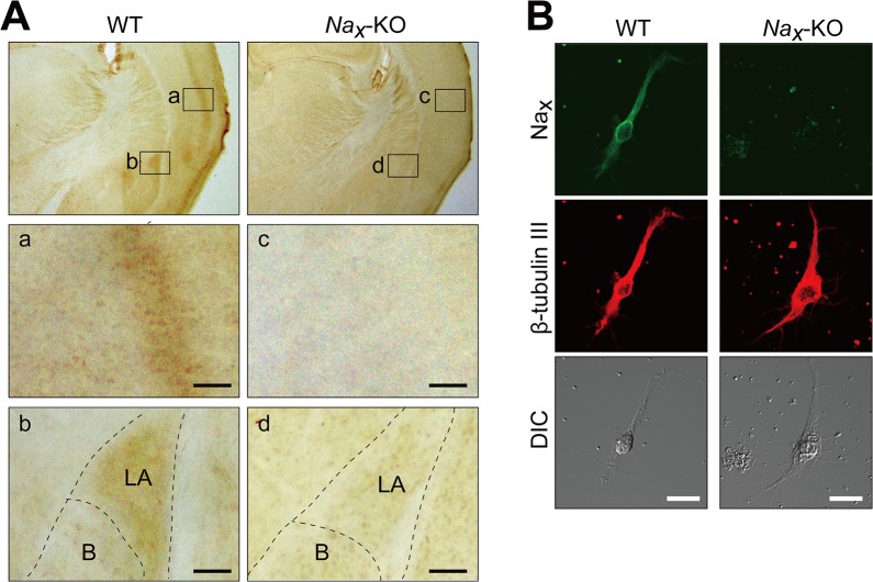
Fig 1. Lateral amygdala neurons express Nax channels.
(A) Immunohistochemical staining of the coronal sections of adult wild-type (WT) and Na x-knockout (Na x-KO) mice with an anti-mNax antibody. The lower panels are magnified views of the square areas inside the upper panel. Immunohistochemical brown staining was observed in the cortex (a) and lateral amygdala (b) in WT mice, but not in Na x-KO mice (c and d). LA, Lateral amygdala; B, Basal amygdala. Scale bars, 50 μm. (B) Double immunofluorescence staining of primary cultured cells obtained from the lateral amygdala of WT and Na x-KO mice with anti-mNax (green) and anti-β-tubulin III (red, a neuronal marker) antibodies. DIC, differential interference contrast image. Scale bars, 20 μm.

