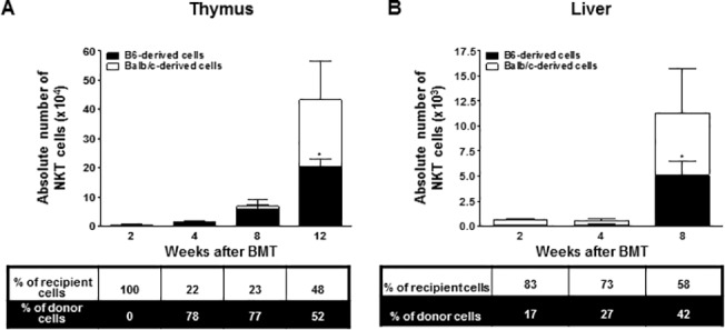
Fig 5. At weeks 2, 4, 8, and 12 after BMT, leukocytes isolated from the thymus (A) and liver (B) were stained with αGalCer-loaded CD1d dimer or anti-TCRβ,-Kb, and-Dd monoclonal antibodies (n = 4 at each time point).

The bars show the ratio and absolute numbers of B6-orginated NKT cells (■) and Balb/c-originated NKT cells (□). Data are shown as the mean ± SD. p values compared to donor-derived cells one week after BMT. *, p ≤ 0.05.
