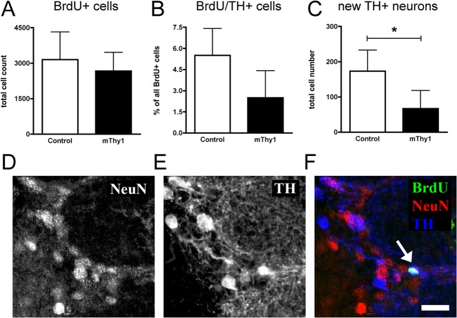
Fig 2. Stereological quantification in the olfactory bulb glomerular cell layer.
the number of BrdU-positive cells did not differ between transgenic a-Synuclein mThy1 and control animals (panel A). The analysis of the percentage of BrdU/TH-double-positive cells revealed a non-significant (p = 0.07) trend (panel B). The numbers of newly generated dopaminergic neurons are significantly reduced in the a-Synuclein transgenic animals (p = 0.04, panel C). Immunofluorescent confocal images of the glomerular cell layer show colocalization of markers of neuronal (NeuN, panel D) and dopaminergic differentiation (TH, panel E) with BrdU, a marker of cell survival (panel F). Scale bar 20μm.

