
Figure 1.
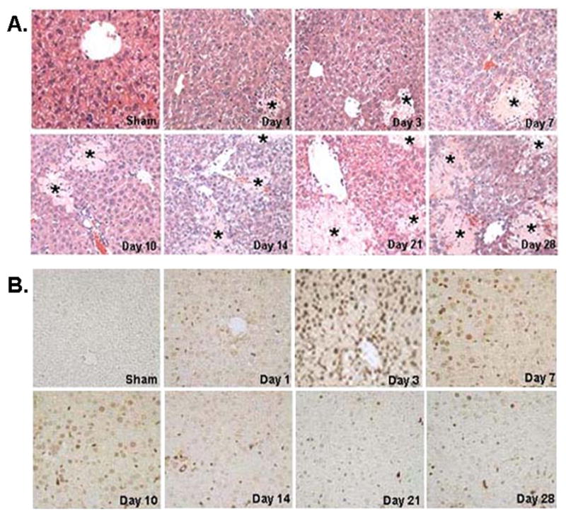
A) Representative liver sections stained with H&E from CBDL and sham-operated mice, day 1 to day 28. The liver was histologically normal in Sham-operated mice. Liver tissues from day 1 on had increased necrosis in CBDL group. Stars indicate areas of hepatic necrosis (H&E staining, 200X). B) Representative liver sections stained with TUNEL from CBDL and sham-operated mice, day 1 to day 28. Cells negative for apoptosis exhibited no nuclear staining, whereas apoptotic cells exhibited brown nuclei staining. Hepatocyte apoptosis was not detected in sham-operated mice. Apoptotic cells were observed on day 1, peaked at day 3, maintained a persistent high level of apoptosis up to day 10, and decreased from day 14 to day 28 (TUNEL staining, 200X).