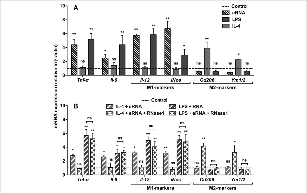
Figure 5. eRNA treatment alters macrophage polarisation markers and pro-inflammatory mediators in BMDM.
A) BMDM were differentiated in the presence of M-CSF-containing L929-conditioned medium and exposed to medium only (Control, dotted line) or to LPS (100 ng/ml), IL-4 (10 ng/ml), or eRNA (10 µg/ml) for 24 h as indicated. B) BMDM were differentiated in the presence of M-CSF-containing L929-conditioned medium and exposed to medium only (Control, dotted line) or to combinations of IL-4 + eRNA and LPS + eRNA in the absence or presence of 1 µg/ml RNase1 (to obtain hydrolysed eRNA) for 24 h as indicated. In all cases, mRNA expression of Tnf-α, Il-6, Il-12, iNos, Cd206 and Ym1/2 was analysed by real-time PCR. Data are expressed as changes in the ratio between target gene and β-actin mRNA expression. The results were obtained from six independent experiments carried out in duplicates. Values represent mean ± SD; ns=non-significant, *p<0.05, **p<0.01 vs control.

