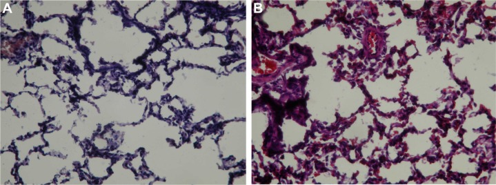
Figure 6.

Pathological examination of lung tissues.
Notes: (A) Group 1 rabbits showed interstitial inflammatory cell infiltrates and pneumocyte hyperplasia (hematoxylin–eosin staining, 100× magnification). (B) Group 2 rabbits showed a pattern very similar to that observed in Group 1 (hematoxylin–eosin staining, 100× magnification). Group 1 was treated with the penicillin-eluting catheter (20 mg/catheter), and Group 2 received systemic intramuscular administration of penicillin (10 mg/kg every 2 days with a non-drug eluting catheter).
