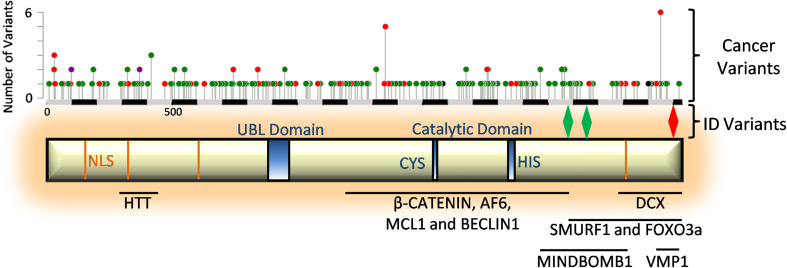
Fig. 1.
Structural information of USP9X. Schematic of USP9X structure showing functional domains and nuclear localisation sequence (NLS) motifs. Below the schematic are the regions of USP9X known to facilitate binding to the listed interacting proteins. Above the schematic is a scale (in amino acids), the localisation of variants associated with ID18 and a histogram of variants found in cancer samples (cBioportal). Red indicates nonsense variants, green represents missense variants and purple indicates both

