
Fig. 3.
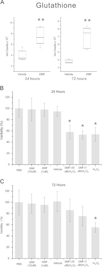
DMF induces glutathione and protects oligodendroglial cells from oxidative stress. (A) Box and whisker plot of GSH levels after 24 and 72 h of treatment with 10 µM DMF. GSH is significantly increased at both time points (**q<0.01, p=0.0006, q=0.004 at 24 h and p=0.003, q=0.004 at 72 h). (B) MTT assay of MO3.13 cells treated with DMF for 24 h and exposed to 400 µM hydrogen peroxide for 2 h. Hydrogen peroxide-treated cells show a significant loss of viability compared to vehicle controls and DMF treatment at both 1 and 10 µM did not significantly alter this reduction in viability. (C) MTT assay of MO3.13 cells treated with DMF for 72 h and challenged with hydrogen peroxide. In contrast to vehicle-treated controls, hydrogen peroxide-treated cells show reduced viability that was rescued by pre-treatment with DMF. *p-value <0.05, Kruskal–Wallis test, N=12 replicates per group, experiment repeated 3 times.