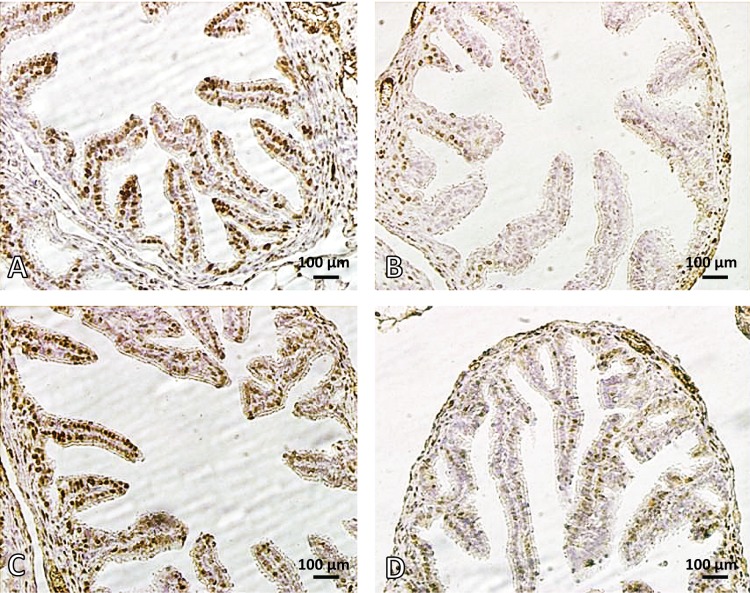
Figure 4.
Photomicrograph of mouse ampulla (oviduct). A: control, B: nicotine treated mouse, C: melatonin treated mouse and D: melatonin+nicotine treated mouse. Brown cells show ER alpha positive cells. Note the reduced numbers of ER alpha positive cells and mucosal folds (MF) in nicotine (B) compared with control (A). In D, melatonin ameliorated the mucosal folds of the oviduct and increased ER alpha. ER alpha Immunostaining. Magnification 400×.

