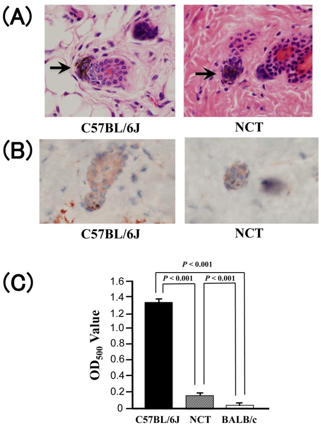
Fig. 2.

(A) Hematoxylin and eosin staining of the dorsal dermis of 12-week-old C57BL/6J and NCT mice (original magnification ×400). Melanin granules in hair bulb are indicated by arrows. (B) Immunohistochemical staining of TRP1 in the dorsal dermis of 12-week-old C57BL/6J and NCT mice (original magnification ×400). (C) Comparison of hair melanin levels between 12-week-old C57BL/6J, NCT, and BALB/c (mean ± SD; n = 3).
