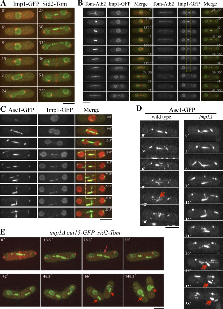
Figure 2.
Imp1 regulates spindle disassembly at the midzone. (A) Time-lapse fluorescence images of Imp1-GFP– and Sid2-Tom–expressing cells. During anaphase B, Imp1-GFP localizes at the central internuclear bridge (dashed squares at 27 and 30 min). (B) Time-lapse fluorescence images of a representative cell expressing Imp1-GFP and the microtubule marker Tom-Atb2. Imp1-GFP colocalizes with the spindle midzone, where antiparallel microtubules interdigitate (dashed area). (C) Colocalization of Imp1-Tom and the spindle midzone marker Ase1-GFP by time-lapse fluorescence microscopy (merge, dashed area). (D) Localization of Ase1-GFP by time-lapse fluorescence in wild-type and imp1Δ cells. Arrows indicate Ase1-GFP disappearance from spindle midzone during normal spindle disassembly in wild-type cells and Ase1-GFP persistence at the midzone domain in imp1Δ cells. (E) Time-lapse fluorescence images of imp1Δ cells coexpressing Cut15-GFP and Sid2-Tom, showing assembled midzone fragments after mitosis (arrows). Bars, 5 µm.

