Figure 3.

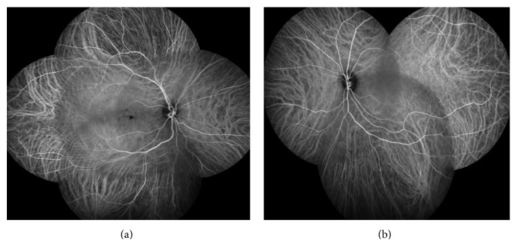
Indocyanine green angiography (ICG) revealed hypofluorescent lesions in both early and late phases, more evident in the RE.

Indocyanine green angiography (ICG) revealed hypofluorescent lesions in both early and late phases, more evident in the RE.