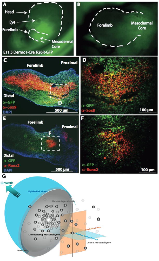
Figure 1. Key extrinsic factors and their role in mesenchyme condensations, e.g. in the murine embryonic limbud at E11.5.
A–F: The ectoderm and mesenchmal core in limbbuds immunostained with antibodies against GFP for Dermo1 lineage (green), Sox9 (red: C,D), and Runx2 (red: E,F). D,F correspond to a high magnification enlargement of the respective dashed areas in the mesodermal core of C,E. In C–F, cells nuclei appear blue due to DAPI staining. G represents the bulbous limb bud schematically, demonstrating the spatial dependence of inductive biochemical cues from the ectoderm (depicted as “epithelial sheet”) as well as their interplay with biophysical cues deriving from the condensation as well as proximity to the ectoderm (epithelial sheet). Refer to 4. Genetic… for further details of gene activity associated with the condensation event. Fig. 1A–F, adapted from [Falls 2007]

