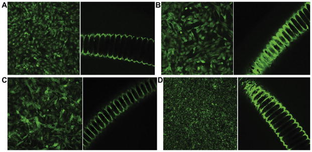
Figure 3.

Control (left) and experimental (right) immunohistochemical staining for α1 integrin (A), α2 integrin (B), β1 integrin (C), and platelet endothelial cell adhesion molecule (D) showing endothelial cell localization on coils. Control images represent staining of cells attached to the surface of the wells rather than the coil segments.
