
Figure 6.
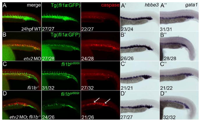
Apoptosis of vascular endothelial cells is expanded in double Etv2;Fli1b-deficient embryos. A-D, Whole-mount immunohistochemical staining of 48-hpf embryos for caspase 3 and green fluorescent protein (GFP) expression. Lateral views shown with anterior to the left. No distinct zones of apoptosis were noted in wild-type (A) and fli1b-/- embryos (C). By contrast, etv2 morphant embryos showed a narrow zone of apoptosis in the vascular plexus (B). D, This zone of apoptosis was expanded in embryos deficient in both fli1b and etv2 (arrows). Hbbe3 (A′-D′) and gata1 (A″-D″) levels determined by whole-mount in situ hybridization show normal erythrocyte staining in all groups indicating the observed apoptosis was not because of a loss of blood cells. Fractions indicate the number of embryos with the observed pattern of marker expression (numerator) and the total evaluated (denominator).